Journal of Clinical and Biomedical Sciences
DOI: 10.58739/jcbs/v14i2.24.36
Year: 2024, Volume: 14, Issue: 2, Pages: 49-55
Original Article
Poorni Bharathi T1, Marla Nisha J2,∗
1 Junior Resident, Department of Pathology, Father Muller Medical College, Kankanady, Mangaluru, Karnataka, India
2 Professor, Department of Pathology, Father Muller Medical College, Kankanady, Mangaluru, Karnataka, India
Received Date:21 May 2024, Accepted Date:13 July 2024, Published Date:23 July 2024
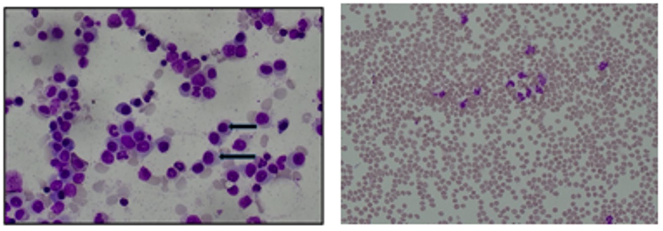
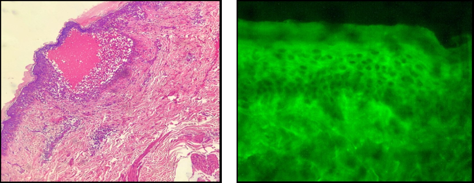
Introduction: Cutaneous noninfectious autoimmune vesiculobullous diseases are a wide variety of lesion. They can show clinical and histopathological overlap, posing diagnostic challenges. Clinically, vesiculobullous lesions might not present with typical morphology and distribution in all cases. The specific histomorphological changes are observed only in an early and intact bulla. Immunofluorescence (IF) is a histochemical staining technique which detects antibodies in the tissue and body fluids. IF an adjuvant to histopathology is considered as gold standard in the diagnosis of vesiculobullous diseases. Methodology: A two-year retrospective cross-sectional descriptive study was conducted in the department of pathology, in a tertiary care center from January 2018 to December 2019. Paraffin embedded hematoxylin and eosin slides were retrieved from the archives and analyzed. Biopsies received in Michel’s medium were utilized for immunofluorescence study. Results: A total of 67 cases with Vesiculobullous lesions were analysed. The most common lesion diagnosed was Bullous Pemphigoid (BP) (29 cases -46.7%), followed by Pemphigus Vulgaris (PV) (19 cases- 30.6%). The highest number of cases were noted in the 7th decade, with a slight female preponderance. Sixteen out of 19 cases of pemphigus vulgaris and 28 out of 29 cases of bullous pemphigoid showed positive histomorphological correlation with IF. The PPV for pemphigus vulgaris was 100% and sensitivity was 84.2%, and in bullous pemphigoid, both the PPV and sensitivity was 96.5%. Higher concordance was noted between Histopathology and IF diagnosis in cases of BP in comparison to PV. Conclusion: Vesiculobullous lesions are heterogeneous in nature and can have clinical and histopathological overlap, posing diagnostic challenges. IF is considered gold standard and is necessary for the diagnosis in every case. A combined clinical, histopathological and IF examination is the best approach to accurate diagnosis.
Keywords
Bullous, Pemphigus, Vesiculobullous, Direct Immunofluorescence
This is an open-access article distributed under the terms of the Creative Commons Attribution License, which permits unrestricted use, distribution, and reproduction in any medium, provided the original author and source are credited.
Published By Sri Devaraj Urs Academy of Higher Education, Kolar, Karnataka
Subscribe now for latest articles and news.