

Journal of Clinical and Biomedical Sciences
Year: 2022, Volume: 12, Issue: 4, Pages: 142-146
Original Article
Revanth R B1,*, Harini Bopaiah2, Anil Kumar Sakalecha3, Yashas Ullas4, Suraj H S4, Rishi Prajwal1
1 Postgraduate, Department of Radio-diagnosis, SDUMC, Kolar, Karnataka, India
2 Associate Professor, Department of Radio-diagnosis, SDUMC, Kolar, Karnataka, India
3 Professor & HOD, Department of Radio-diagnosis, SDUMC, Kolar, Karnataka, India
4 Senior Resident, Department of Radio-diagnosis, SDUMC, Kolar, Karnataka, India
*Corresponding author email: [email protected]
Received Date:20 October 2022, Accepted Date:30 November 2022, Published Date:22 December 2022
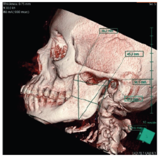
Background: The ability to identify sex is essential for identification and plays a significant role in forensic medicine and medico legal investigations. Identification's first purpose is sex determination, which is followed by estimations of age, size, and ethnic population, all of which are influenced by the gender. When identifying morphologic characteristics for gender determination, gender analysis and estimate are carried out with almost 90% to 100% accuracy using a whole skeleton. On the other hand, partial or fragmented bones and badly injured bodies, such as in the case of a mass tragedy, might be challenging to analyse. The skull is the second-best marker for gender identification after the pelvis. However, as the mandible is the biggest bone of the skull, it may be crucial in circumstances when a whole skull is not recovered for gender identification. Objectives: To ascertain how computed tomography can be utilised to know a person's gender with the help of mandibular measurements. Methods: The study was done with the Siemens Somatom 16 slice CT machine. Patients who match the inclusion criteria and have received CT for a wide range of reasons will be considered. Various measurements such as Condylar height, Maximum ramus breadth, Minimum ramus breadth projective height of ramus, Coronoid height shall be measured. Data analysis must be done with SPSS 20. For comparisons between the right and left sides, the student t-test shall be applied. Results: 58 individuals between the ages of 18 and 35 (29 females, mean age, 26.3, 29 males, men age, 26.9 & overall mean for both sexes was 26.6 years). In the present study, mean score of five distinct mandibular parameters were calculated. All mandibular ramus variables on CT models were discovered to exhibit a statistically significant gender difference (p = 0.001). It was discovered that the mean condylar height was 64.5 mm in men and 59.56 mm in women, which was a significant (P = 0.001). It was noteworthy (P = 0.001) that whereas the mean maximal ramus width was 35.0 mm in females and 38.0 mm in men. Males and females had different mean values for the minimum ramus width, which were 31.3 mm and 28.8 mm, respectively (P = 0.001). Males and females had mean ramus projection height measurements of 52.38 mm and 47.91 mm, respectively. This difference was significant (P = 0.004). The mean coronoid height was 63.42 mm in men and 56.80 mm in women, which was significant (P = 0.001). 27 out of 29 female mandibular measures correctly identified the gender, with a prediction rate of 96.4%, and 27 out of 29 male mandibular measurements correctly identified the gender, with a prediction rate of 96.4%. The threshold for determining a per- son's sex. As a cut-off for predicting male sex, values over 62.8 mm for condylar height, 36.9 mm for maximum ramus height, 30.0 mm for lowest ramus height, 50.4 mm for projective ramus height, and 61.3 mm for coronoid height can be used. Conclusion: In forensic medicine, the assessment of mandible, particularly the ramus area, plays a crucial factor in determining gender. Future research should evaluate gender determination recommendations using various criteria in bigger sample sizes and across a range of age groups. Although there are differences in ramus measurements between both the genders, there are variations in measures between societies. Therefore, to accurately identify gender from skeletal remains, criteria that are unique to each population must be developed. We think the results of this study will be helpful to radiologists making diagnoses and surgeons working on the mandible and facial area.
Keywords: Mandible, Determination of gender, Paranasal sinuses
This is an open-access article distributed under the terms of the Creative Commons Attribution License, which permits unrestricted use, distribution, and reproduction in any medium, provided the original author and source are credited.
Published By Sri Devaraj Urs Academy of Higher Education, Kolar, Karnataka
Subscribe now for latest articles and news.