Journal of Clinical and Biomedical Sciences
DOI: 10.58739/jcbs/v16i1.25.247
Year: 2026, Volume: 16, Issue: 1, Pages: 40-45
Original Article
Manish Kotwani1*, Deepti Kotwani1, Swapnil Lemle2
1Associate Professor, Department of Anaesthesiology, Lokmanya Tilak Municipal Medical College, Sion, Mumbai - 400022, Maharashtra, India.
2Senior Resident, Department of Anaesthesiology, Lokmanya Tilak Municipal Medical College, Sion, Mumbai - 400022, Maharashtra, India.
*Corresponding Author
Email: [email protected]
Received Date:27 May 2025, Accepted Date:27 September 2025, Published Date:19 April 2026
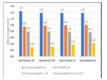
Background: Ultrasound-guided internal jugular vein (IJV) cannulation is standard practice, but the risk of accidental common carotid artery (CCA) puncture persists, especially in obese patients. Head rotation manoeuvres aim to improve IJV accessibility but may increase IJV-CCA overlap. The optimal degree of head rotation to minimise this overlap although studied in normal patients remains largely underexplored in obese patients. Aims: To determine the head rotation angle (15°–60°) that maximizes IJV diameter while minimizing IJV-CCA overlap in obese patients (BMI ≥25 kg/m²). Material and Methods: Sixty obese patients were stratified by BMI (Group 1: 25–29.9; Group 2: 30–39.9; Group 3: ≥40) in the pre-operative room. In supine position with 15° Trendelenburg tilt, right IJV and CCA were imaged using 5–12 MHz linear probe at cricoid level. Transverse and anteroposterior diameters of the right sided IJV and CCA and their overlap were measured using real time ultrasound at 15°, 30°, 45° and 60° of head rotations. Results: IJV-CCA overlap increased significantly with rotation: 34.9% (with15°) to 55.3% (with 60°) (P<0.001). Overlap was highest in Group 3 (BMI ≥40). No significant change in IJV diameter was observed. The safest rotation was ≤45° for BMI 30–39.9 and ≤60° for BMI 25–29.9. Conclusion: Head rotation >45° in obese patients significantly increases CCA puncture risk. Ultrasound-guided cannulation at ≤45° is recommended for BMI ≥30 kg/m².
Keywords: Internal jugular vein, Obesity, Ultrasound guidance, Head rotation, Common carotid artery
1. Comparative Sonoanatomy of Classic “Short Axis” Probe Position with a Novel “Medial-oblique” Probe Position for Ultrasound-guided Internal Jugular Vein Cannulation: A Crossover Study. The Journal of Emergency Medicine. 2015; 48 (5). Available from: https://doi.org/10.1016/j.jemermed.2014.07.062
2. Central venous access sites for the prevention of venous thrombosis, stenosis and infection. Cochrane Database of Systematic Reviews. 2012; 2012 (3). Available from: https://doi.org/10.1002/14651858.cd004084.pub3
3. Guidelines for Performing Ultrasound Guided Vascular Cannulation. Anesthesia & Analgesia. 2012; 114 (1). Available from: https://doi.org/10.1213/ane.0b013e3182407cd8
4. Is Head Rotation Preferred During Right Internal Jugular Vein Cannulation in Obese Asians?. Journal of Anesthesia & Clinical Research. 2012; 03 (10). Available from: https://doi.org/10.4172/2155-6148.1000245
5. Iatrogenesis Imperfecta: Stroke caused by Accidental Carotid Artery Catheterization. The Journal of Vascular Access. 2014; 15 (6). Available from: https://doi.org/10.5301/jva.5000246
6. Arterial trauma during central venous catheter insertion: Case series, review and proposed algorithm. Journal of Vascular Surgery. 2008; 48 (4). Available from: https://doi.org/10.1016/j.jvs.2008.04.046
7. Rajinikanth J, Stephen E, Agarwal S. Complication of central venous cannulation. Canadian Journal of Surgery.2008 Oct;51(5):E113–E114
8. Sono-anatomical analysis of right internal jugular vein and carotid artery at different levels of positive end-expiratory pressure in anaesthetised paralysed patients. Indian Journal of Anaesthesia. 2018; 62 (4). Available from: https://doi.org/10.4103/ija.ija_716_17
9. Anatomical relationship between the common carotid artery and the internal jugular vein during head rotation. Ultrasound. 2014; 22 (2). Available from: https://doi.org/10.1177/1742271x14524571
10. The Impact of Trendelenburg Position and Positive End-Expiratory Pressure on the Internal Jugular Cross-Sectional Area. Anesthesia & Analgesia. 2010; 111 (2). Available from: https://doi.org/10.1213/ane.0b013e3181e2fe41
11. The Effects of the Simulated Valsalva Maneuver, Liver Compression, and/or Trendelenburg Position on the Cross-Sectional Area of the Internal Jugular Vein in Infants and Young Children. Anesthesia & Analgesia. 2002; 94 (2). Available from: https://doi.org/10.1213/00000539-200202000-00004
12. Effects of four different positive airway pressures on right internal jugular vein catheterisation. European Journal of Anaesthesiology. 2012; 29 (5). Available from: https://doi.org/10.1097/eja.0b013e32834f23a3
13. Head Rotation During Internal Jugular Vein Cannulation and the Risk of Carotid Artery Puncture. Anesthesia & Analgesia. 1996; 82 (1). Available from: https://doi.org/10.1097/00000539-199601000-00022
14. Is a Neutral Head Position Safer than 45-Degree Neck Rotation During Ultrasound-Guided Internal Jugular Vein Cannulation? Results of a Randomized Controlled Clinical Trial. Anesthesia & Analgesia. 2012; 114 (4). Available from: https://doi.org/10.1213/ane.0b013e3182459917
15. The Effectiveness Of Trendelenburg Positioning On The Cross-Sectional Area Of The Right Internal Jugular Vein In Obese Patients. Pakistan Journal of Medical Sciences. 1969; 31 (4). Available from: https://doi.org/10.12669/pjms.314.7326
16. Sibai AN, Loutfi E, Itani M, Baraka A. Ultrasound evaluation of the anatomical characteristics of the internal jugular vein and carotid artery – facilitation of internal jugular vein cannulation. Middle East Journal of Anesthesiology. 2008;19:1305‑20.
17. A comparison of internal jugular vein cannulation by ultrasound-guided and anatomical landmark technique in resource-limited emergency department setting. Journal of Medical Ultrasound. 2019; 27 (4). Available from: https://doi.org/10.4103/jmu.jmu_2_19
18. Optimal Head Rotation for Internal Jugular Vein Cannulation When Relying on External Landmarks. Anesthesia & Analgesia. 2004; 99 (4). Available from: https://doi.org/10.1213/01.ane.0000132908.77111.ca
19. Assessment of head and neck position for optimal ultrasonographic visualisation of the internal jugular vein and its relation to the common carotid artery: a prospective observational study. Journal of Anaesthesiology Clinical Pharmacology. 2020; 36 (1). Available from: https://doi.org/10.4103/joacp.joacp_330_18
20. Umaña M, García A, Bustamante L, Castillo JL, Sebastián Martínez J. Variations in the anatomical relationship between the common carotid artery and the internal jugular vein: an ultrasonographic study. Colombia Médica. 2015;46(2):54–9.
21. An unseen danger: Frequency of posterior vessel wall penetration by needles during attempts to place internal jugular vein central catheters using ultrasound guidance*. Critical Care Medicine. 2009; 37 (8). Available from: https://doi.org/10.1097/ccm.0b013e3181a067d4
This is an open-access article distributed under the terms of the Creative Commons Attribution License, which permits unrestricted use, distribution, and reproduction in any medium, provided the original author and source are credited.
Published By Sri Devaraj Urs Academy of Higher Education, Kolar, Karnataka
Subscribe now for latest articles and news.