Journal of Clinical and Biomedical Sciences
Year: 2022, Volume: 12, Issue: 4, Pages: 147-150
Case Report
Shilpa M D1,*, Harendrakumar M L2, Dave Prakash3
Received Date:28 August 2022, Accepted Date:09 September 2022, Published Date:25 December 2022
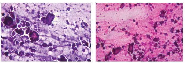
Calcinosis cutis also commonly called as Calciphylaxis is one of the uncommon conditions characterized by precipitation and deposition of phosphate and calcium salts in the dermis and subcutaneous tissues. We report two different cases clinical diagnosed as malignancy and final diagnosis was made on FNAC which correlated with the histopathology. A 45-year-old women came with a compliant of swelling over lateral aspect of thigh on left side since 6 months. Provisional diagnosis of liposarcoma was made clinically and on FNAC given as Calcinosis cutis which correlated with histopathology. A 62-year-old women came with compliant of swelling over lateral aspect of thigh on right side since 10 months associated with pain. Provisional diagnosis of soft tissue sarcoma was made clinically and advised for FNAC clinically and on FNAC given as Calcinosis cutis which correlated with histopathology. Calcinosis cutis is a condition in which there is organized and localized deposition of calcium in the skin. The evolution of the lesions depends on the aetiology of the calcification. The pathogenesis still remains unclear, but several theories have been suggested for this. Calcinosis cutis is a potential mimic of a neoplastic lesion. FNAC is a simple, rapid and reliable technique and which helps in diagnosis of such lesions.
Keywords: Mimicker, Fine needle aspiration cytology, Diagnosis
This is an open-access article distributed under the terms of the Creative Commons Attribution License, which permits unrestricted use, distribution, and reproduction in any medium, provided the original author and source are credited.
Published By Sri Devaraj Urs Academy of Higher Education, Kolar, Karnataka
Subscribe now for latest articles and news.