
JOURNAL OF CLINICAL AND BIOMEDICAL SCIENCES

Article

Journal of Clinical and Biomedical Sciences

Year: 2024, Volume: 14, Issue: 4, Pages: 121-128

Review Article

The Role of Artificial Intelligence in Enhancing Diabetic Retinopathy Lesion Detection: A Review

Received Date:07 August 2024, Accepted Date:01 October 2024, Published Date:20 December 2024

Abstract

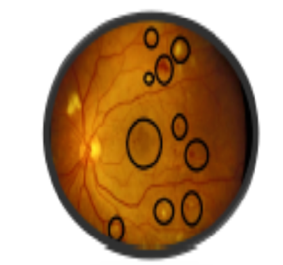
Diabetic retinopathy represents a significant microvascular complication associated with prolonged diabetes mellitus and serves as a leading cause of blindness, particularly in developing nations. For the patient's vision to be adequately preserved, early identification of DR is essential. In order to treat the disease, the patient must maintain his or her current level of vision since the disease is irreversible. The Clinical diagnosis demands significant time and the specialized knowledge of an experienced ophthalmologist and also identifying the disease features in images is also more challenging, particularly in the early stages of the disease when disease features are less noticeable. Therefore, deep learning algorithms have been used for the early diagnosis of DR in recent years, and medical image analysis utilising machine learning has demonstrated to be effective in evaluating retinal fundus images. This review's objective is to go over the numerous Deep learning techniques for automated computer-aided analysis of microaneurysms, haemorrhages, and exudates were also addressed, along with a knowledge gap in DR identification. As part of future research, this review seeks to systematize the available algorithms for ease of use and guidance by researchers.

Keywords: Diabetic Retinopathy Review, Microaneurysms, Haemorrhages, Exudates, Red Lesions, Deep Learning

References

  1. Sen M, Honavar SG. Eye Care for All. Indian Journal of Ophthalmology. 2022;70(9):3169–3170. Available from: https://journals.lww.com/ijo/fulltext/2022/09000/eye_care_for_all.1.aspx
  2. Ting DSW, Cheung GCM, Wong TY. Diabetic retinopathy: global prevalence, major risk factors, screening practices and public health challenges: a review. Clinical and Experimental Ophthalmology. 2016;44(4):260–277. Available from: https://doi.org/10.1111/ceo.12696
  3. Abbas Q, Fondon I, Sarmiento A, Jiménez S, Alemany P. Automatic recognition of severity level for diagnosis of DR using deep visual features. Medical & Biological Engineering & Computing. 2017;55(11):1959–1974. Available from: https://doi.org/10.1007/s11517-017-1638-6
  4. Li Z, Keel S, Liu C, He Y, Meng W, Scheetz J, et al. An automated grading system for detection of vision-threatening referable DR on the basis of colour fundus photographs. Diabetes care. 2018;41(12):2509–2516. Available from: https://doi.org/10.2337/dc18-0147
  5. Tajbakhsh N, Shin JY, Gurudu SR, Hurst RT, Kendall CB, Gotway MB, et al. Convolutional neural networks for medical image analysis: Full training or fine tuning. IEEE transactions on medical imaging. 2016;35(5):1299–1312. Available from: https://doi.org/10.1109/TMI.2016.2535302
  6. Erhan D, Bengio Y, Courville A, Manzagol PA, Vincent P, Bengio S. Why does unsupervised pre-training help deep learning. Journal of Machine Learning Research. 2010;11:625–660. Available from: https://www.jmlr.org/papers/volume11/erhan10a/erhan10a.pdf
  7. Updated 2017 ICO guidelines for diabetic eye care. San Francisco (CA): International Council of Ophthalmology, 1–33 . Available from: http://www.icoph.org/downloads/ ICOGuidelinesforDiabeticEyeCare.pdf
  8. Long S, Chen J, Hu A, Liu H, Chen Z, Zheng D. Microaneurysms detection in color fundus images using machine learning based on directional local contrast. Biomedical engineering online. 2020;19(1):1–23. Available from: https://doi.org/10.1186/s12938-020-00766-3
  9. Liao Y, Xia H, Song S, Li H. Microaneurysm detection in fundus images based on a novel end-to-end convolutional neural network. Biocybernetics and Biomedical Engineering. 2021;41(2):589–604. Available from: https://doi.org/10.1016/j.bbe.2021.04.005
  10. Mateen M, Malik TS, Hayat S, Hameed M, Sun S, Wen J. Deep Learning Approach for Automatic Microaneurysms Detection. Sensors. 2022;22(2):1–14. Available from: https://doi.org/10.3390/s22020542
  11. Wu J, Zhang S, Xiao Z, Zhang F, Geng L, Lou S, et al. Hemorrhage detection in fundus image based on 2D Gaussian fitting and human visual characteristics. Optics & Laser Technology. 2019;110:69–77. Available from: https://doi.org/10.1016/j.optlastec.2018.07.049
  12. Prentašić P, Lončarić S. Detection of exudates in fundus photographs using deep neural networks and anatomical landmark detection fusion. Computer Methods and Programs in Biomedicine. 2016;137:281–292. Available from: https://doi.org/10.1016/j.cmpb.2016.09.018
  13. Zago GT, Andreão RV, Dorizzi B, Salles EOT. Diabetic retinopathy detection using red lesion localization and convolutional neural networks. Computers in Biology and Medicine. 2020;116. Available from: https://doi.org/10.1016/j.compbiomed.2019.103537
  14. Gulshan V, Peng L, Coram M, Stumpe MC, Wu D, Narayanaswamy A, et al. Development and Validation of a Deep Learning Algorithm for Detection of Diabetic Retinopathy in Retinal Fundus Photographs. JAMA. 2016;316(22):2402–2410. Available from: https://doi.org/10.1001/jama.2016.17216
  15. Grzybowski A, Brona P, Lim G, Ruamviboonsuk P, Tan GS, Abramoff M, et al. Artificial intelligence for diabetic retinopathy screening. Eye . 2020;34:451–460. Available from: https://doi.org/10.1038/s41433-019-0566-0
  16. Kanagasingam Y, Xiao D, Vignarajan J, Preetham A, Tay-Kearney ML, Mehrotra A. Evaluation of Artificial Intelligence–Based Grading of Diabetic Retinopathy in Primary Care. JAMA Network Open. 2018;1(5). Available from: https://dx.doi.org/10.1001/jamanetworkopen.2018.2665
  17. Abràmoff MD, Lou Y, Erginay A, Clarida W, Amelon R, Folk JC, et al. Improved Automated Detection of Diabetic Retinopathy on a Publicly Available Dataset Through Integration of Deep Learning. Investigative Opthalmology & Visual Science. 2016;57(13). Available from: https://dx.doi.org/10.1167/iovs.16-19964
  18. Gulshan V, Rajan RP, Widner K, Wu D, Wubbels P, Rhodes T, et al. Performance of a Deep-Learning Algorithm vs Manual Grading for Detecting Diabetic Retinopathy in India. JAMA Ophthalmology. 2019;137(9). Available from: https://dx.doi.org/10.1001/jamaophthalmol.2019.2004
  19. Kiyota N, Shiga Y, Omodaka K, Pak K, Nakazawa T. Time-Course Changes in Optic Nerve Head Blood Flow and Retinal Nerve Fiber Layer Thickness in Eyes with Open-angle Glaucoma. Ophthalmology. 2021;128(5):663–671. Available from: https://dx.doi.org/10.1016/j.ophtha.2020.10.010
  20. Chandrasekaran PR, Madanagopalan VG, Narayanan R. DR in pregnancy - A review. Indian Journal of Ophthalmology. 2021;69(11):3015–3025. Available from: http://dx.doi.org/10.4103/ijo.IJO_1377_21
  21. Wild SH, Roglic G, Green A, Sicree R, King H. Global Prevalence of Diabetes: Estimates for the Year 2000 and Projections for 2030. Diabetes Care. 2004;27(10). Available from: https://dx.doi.org/10.2337/diacare.27.10.2569-a

Copyright

This is an open-access article distributed under the terms of the Creative Commons Attribution License, which permits unrestricted use, distribution, and reproduction in any medium, provided the original author and source are credited.

Published By Sri Devaraj Urs Academy of Higher Education, Kolar, Karnataka

DON'T MISS OUT!

Subscribe now for latest articles and news.