Journal of Clinical and Biomedical Sciences
DOI: 10.58739/jcbs/v16i1.25.339
Year: 2026, Volume: 16, Issue: 1, Pages: 81-83
Case Report
Nellaiappan Chelliah1, Thangameena Muthukumar2, Naveen S3*
1Professor, Barnard Institute of Radiology, Madras Medical College, Chennai 600003, Tamil Nadu, India.
2Assistant Professor, Barnard Institute of Radiology, Madras Medical College, Chennai 600003, Tamil Nadu, India.
3Final year Resident, Barnard Institute of Radiology, Madras Medical College, Chennai 600003, Tamil Nadu, India.
*Corresponding Author
Email: [email protected]
Received Date:05 September 2025, Accepted Date:20 January 2026, Published Date:21 April 2026
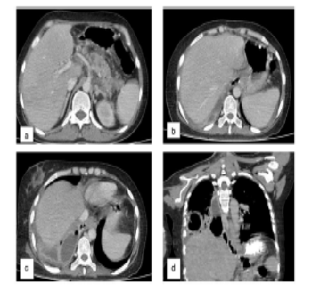
Acute pancreatitis is an inflammatory condition with a wide clinical spectrum, ranging from mild interstitial involvement to severe necrotizing disease. Imaging plays a vital role in identifying complications, assessing severity, and guiding timely clinical decisions. This study presents a series of three radiologically confirmed cases that illustrate the diverse complications of pancreatitis, including pseudocyst formation, walled-off necrosis, infected necrosis, pseudoaneurysm, splenic vein thrombosis and pancreaticopleural fistula. Cross-sectional imaging, particularly contrast-enhanced CT, was essential in detecting these findings and revealing critical complications such as hemorrhagic transformation, vascular involvement, and thoracic extension. Recognition of these varied imaging patterns is crucial, as complications may mimic other pathologies and significantly influence prognosis and management. A thorough understanding of the radiologic spectrum of pancreatitis-related complications enables accurate diagnosis and effective multidisciplinary care.
Keywords: Acute pancreatitis, Necrotizing pancreatitis, Walled off necrosis, Infected pancreatic necrosis, Pancreatic pseudocyst, Hemorrhagic pseudocyst, Pseudoaneurysm, Contrast enhanced CT, Pancreaticopleural fistula, Splenic vein thrombosis
1. Ranson JH, Balthazar E, Caccavale R, Cooper M. Computed Tomography and the Prediction of Pancreatic Abscess in Acute Pancreatitis. Annals of Surgery. 1985; 201 (5). Available from: https://doi.org/10.1097/00000658-198505000-00016
2. Kim HC, Yang DM, Kim HJ, Lee DH, Ko YT, Lim JW. Computed tomography appearances of various complications associated with pancreatic pseudocysts. Acta Radiologica. 2008; 49 (7). Available from: https://doi.org/10.1080/02841850802104932
3. Grassedonio E, Toia P, La Grutta L, Palmucci S, Smeraldi T, Cutaia G, et al. Role of computed tomography and magnetic resonance imaging in local complications of acute pancreatitis. Gland Surgery. 2019; 8 (2). Available from: https://doi.org/10.21037/gs.2018.12.07
4. Morgan DE. Imaging of Acute Pancreatitis and Its Complications. Clinical Gastroenterology and Hepatology. 2008; 6 (10). Available from: https://doi.org/10.1016/j.cgh.2008.07.012
5. Miller FH. Radiology of the pancreas, gallbladder, and biliary tract. Radiologic Clinics of North America. 2002; 40 (6). Available from: https://doi.org/10.1016/s0033-8389(02)00097-0
6. Chishty IA, Bari V, Pasha S, Burhan D, Haider Z, Rafique Z. (2005). Role of computed tomography in acute pancreatitis and its complications among age groups. Journal of Pakistan Medical Association, 55(10), 431-435.
7. Vriens PW, van de Linde P, Slotema ET, Warmerdam PE, Breslau PJ. Computed Tomography Severity Index Is an Early Prognostic Tool for Acute Pancreatitis. Journal of the American College of Surgeons. 2005; 201 (4). Available from: https://doi.org/10.1016/j.jamcollsurg.2005.06.269
This is an open-access article distributed under the terms of the Creative Commons Attribution License, which permits unrestricted use, distribution, and reproduction in any medium, provided the original author and source are credited.
Published By Sri Devaraj Urs Academy of Higher Education, Kolar, Karnataka
Subscribe now for latest articles and news.