Journal of Clinical and Biomedical Sciences
DOI: 10.58739/jcbs/v16i2.25.326
Year: 2026, Volume: 16, Issue: 2, Pages: 41-46
Original Article
Swathy Shanker1*, Deepak Lavatt2, Swathi Raj3, Ramesh P K 4, Sathi P P 5
1Assistant Professor, Department of PG studies and Research, KMCT Medical College, Kerala, India.
2Assistant Professor, Department of Surgery, KMCT Medical College, Kerala, India.
3Senior Resident, Department of Pathology, KMCT Medical College, Kerala, India.
4Professor, Department of Surgery, KMCT Medical College, Kerala, India.
5Professor, Department of Pathology, KMCT Medical College, Kerala, India.
*Corresponding Author
Email: [email protected]
Received Date:18 August 2025, Accepted Date:14 November 2025, Published Date:15 June 2026
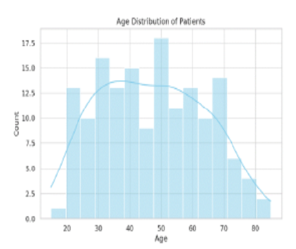
Context: Gallstones are a recurrent hepatobiliary disease seen in around 10-15% of the population. Various classification systems have been proposed for the classification of gall stones, and the widely used system utilizes the morphology and chemical content of gall stones. Aims: In this study we aim to evaluate the incidence of gall stones and to describe the clinicopathological correlation of the disease in the study population. Settings and Design: This is a retrospective, descriptive study done using the medical records of patients presented with cholelithiasis over a period of 2 years. Methods and Material: A total of 155 patients were included in the study. The histopathological diagnoses of the cholecystectomy specimens were retrieved from the archived reports, and the data was evaluated with patient details. Statistical analysis used: Data was entered in Microsoft Excel and analysed for correlation between various variables using SPSS software version 22.0. Results: The ages of the patients varied from 15 years old to 85 years old, with the mean age of the study population being 47 years. A female predominance was seen, and pigment stones were found to be the most common type. Majority of the patients (58.7%) had no coexisting comorbidities while the most common comorbidity seen in patients with pigmented stones was that of diabetes (13.3%). Among the patients with cholesterol stones 15.4% of the patients showed presence of thyroid disorder and two out of the three patients with mixed type of gall stones showed coexisting diabetes. Conclusions: Gall stones were found more commonly among women with an increased incidence in women of the reproductive age group. The most common gall stone found in the study population was that of pigment stones followed by cholesterol stones. Patients with diabetes showed an increased incidence of pigmented and mixed stones while patients with thyroid disorder showed an increased incidence of cholesterol stones. Key Messages: An increased incidence of pigmented and mixed gall stones has been observed among patients with diabetes. A larger, population based study is warranted to evaluate the underlying mechanism.
Keywords: Cholelithiasis, Pigment stones, Cholesterol stones
1. Schwesinger WH, Kurtin WE, Page CP, Stewart RM, Johnson R. Soluble dietary fiber protects against cholesterol gallstone formation. The American Journal of Surgery. 1999; 177 (4). Available from: https://doi.org/10.1016/s0002-9610(99)00047-1
2. Brett M, Barker DJP. The World Distribution of Gallstones. International Journal of Epidemiology. 1976; 5 (4). Available from: https://doi.org/10.1093/ije/5.4.335
3. Shabanzadeh DM. Incidence of gallstone disease and complications. Current Opinion in Gastroenterology. 2018; 34 (2). Available from: https://doi.org/10.1097/mog.0000000000000418
4. Stinton LM, Shaffer EA. Epidemiology of Gallbladder Disease: Cholelithiasis and Cancer. Gut and Liver. 2012; 6 (2). Available from: https://doi.org/10.5009/gnl.2012.6.2.172
5. Schirmer BD, Winters KL, Edlich RF. Cholelithiasis and Cholecystitis. Journal of Long-Term Effects of Medical Implants. 2005; 15 (3). Available from: https://doi.org/10.1615/jlongtermeffmedimplants.v15.i3.90
6. Duane WC. Pathogenesis of Gallstones: Implications for Management. Hospital Practice. 1990; 25 (3). Available from: https://doi.org/10.1080/21548331.1990.11703924
7. Rigas B, Torosis J, McDougall CJ, Vener KJ, Spiro HM. The Circadian Rhythm of Biliary Colic. Journal of Clinical Gastroenterology. 1990; 12 (4). Available from: https://doi.org/10.1097/00004836-199008000-00011
8. Shabanzadeh DM. New determinants for gallstone disease?. Danish Medical Journal. 2018; 65 (2). Available from: https://pubmed.ncbi.nlm.nih.gov/29393043/
9. Trotman BW, Soloway RD. Pigment Gallstone Disease: Summary of the National Institutes of Health—International Workshop. Hepatology. 1982; 2 (6). Available from: https://doi.org/10.1002/hep.1840020624
10. Suzuki N, Sato T. A new classification of gallstone. <I>Journal of Biliary Tract and Pancreas</I> 1986;7:1467-70.
11. Channa NA. Gallstone disease: a review. <I>Pakistan Armed Forces Medical Journal</I>. 2008 Jun 30;58(2):197-208.
12. Shaffer EA. Gallstone disease: Epidemiology of gallbladder stone disease. Best Practice & Research Clinical Gastroenterology. 2006; 20 (6). Available from: https://doi.org/10.1016/j.bpg.2006.05.004
13. Trotman BW, Petrella EJ, Soloway RD, Sanchez HM, Morris TA, Miller WT. Evaluation of Radiographic Lucency or Opaqueness of Gallstones as a Means of Identifying Cholesterol or Pigment Stones. Gastroenterology. 1975; 68 (6). Available from: https://doi.org/10.1016/s0016-5085(75)80145-4
14. Kurtin WE, Schwesinger WH, Diehl AK. Age-related changes in the chemical composition of gallstones. <I>International Journal of Surgical Investigation</I>. 2000;2:299–307.
15. Sharma MA. Towards a safer cholecystectomy-The fundus to porta approach.<I>Indian Journal of Surgery</I>.1997;59(4):141-5.
16. Selvi TR, Sinha P, Subramaniam PM, Konapur PG, Prabha CV. A clinicopathological study of cholecystitis with special reference to analysis of cholelithiasis. <I>International Journal Of Basic Medical Science</I>. 2011;2(2):68-72.
17. Veerabhadrappa PS, Tank P, Singh A, Goel S, Nathwani P. A study of gall stone disease from a tertiary care center of Madhya Pradesh, India. International Surgery Journal. 2017; 4 (2). Available from: https://doi.org/10.18203/2349-2902.isj20170222
18. Wang X, Yu W, Jiang G, Li H, Li S, Xie L, <I>et al</I>. Global Epidemiology of Gallstones in the 21st Century: A Systematic Review and Meta-Analysis. Clinical Gastroenterology and Hepatology. 2024; 22 (8). Available from: https://doi.org/10.1016/j.cgh.2024.01.051
19. Valdivieso V, Covarrubias C, Siegel F, Cruz F. Pregnancy and Cholelithiasis: Pathogenesis and Natural Course of Gallstones Diagnosed in Early Puerperium. Hepatology. 1993; 17 (1). Available from: https://doi.org/10.1002/hep.1840170102
20. Cirillo DJ, Wallace RB, Rodabough RJ, Greenland P, LaCroix AZ, Limacher MC, <I>et al</I>. Effect of Estrogen Therapy on Gallbladder Disease. JAMA. 2005; 293 (3). Available from: https://doi.org/10.1001/jama.293.3.330
21. Qiao T, Ma RH, Luo XB, Yang LQ, Luo ZI, Zheng PM. The Systematic Classification of Gallbladder Stones. PLoS ONE. 2013; 8 (10). Available from: https://doi.org/10.1371/journal.pone.0074887
22. Maskey CP, Shrestha ML, Sato Y. Gallstone in TUTH. <I>JIOM</I>.1990;12:45-54
23. Paumgartner G, Gerok W, Bertolotti M, Bortolotti S, Menozzi D. Ageing and bile acid metabolism: Studies on 7α hydroxylation of cholesterol in humans. In: <I>Paumgartner G, Gerok W, editors. Trends in Bile Acid Research</I>. Lancaster: Kluwer Academic Publishers; 1989: 75-78.
24. Einarsson K, Nilsell K, Leijd B, Angelin B. Influence of Age on Secretion of Cholesterol and Synthesis of Bile Acids by the Liver. New England Journal of Medicine. 1985; 313 (5). Available from: https://doi.org/10.1056/nejm198508013130501
25. Zeng F, Hai H, Jin L. Analysis on Infrared Spectrum of Human Body Gallstone with Comparative Method. <I>Spectroscopy and Spectral Analysis</I>. 2001; 21 (3): 314–316.
26. Cao J, Wu K, Wu Z, Na P, Dai X. Qualitative analysis of gallstones from 472 patients with cholelithiasis from Ningxia using Infrared Spectrum. <I>Journal of Chinese Physician</I>. 2006; 8 (8): 1111–1113.
27. Chandran P, Kuchhal NK, Garg P, Pundir CS. An extended chemical analysis of gallstone. Indian Journal of Clinical Biochemistry. 2007; 22 (2). Available from: https://doi.org/10.1007/bf02913334
28. Sun H, Warren J, Yip J, Ji Y, Hao S, Han W, <I>et al</I>. Factors Influencing Gallstone Formation: A Review of the Literature. Biomolecules. 2022; 12 (4). Available from: https://doi.org/10.3390/biom12040550
29. Kleiner O, Ramesh J, Huleihel M, Cohen B, Kantarovich K, Levi C, <I>et al</I>. A comparative study of gallstones from children and adults using FTIR spectroscopy and fluorescence microscopy. BMC Gastroenterology. 2002; 2 (1). Available from: https://doi.org/10.1186/1471-230x-2-3
30. Lowenfels AB, Lindstrom CG, Conway MJ, Hastings PR. Gallstones and risk of gallbladder cancer. <I>Journal of the National Cancer Institute</I>. 1985;75(1):77–80.
31. Stinton LM, Shaffer EA. Epidemiology of Gallbladder Disease: Cholelithiasis and Cancer. Gut and Liver. 2012; 6 (2). Available from: https://doi.org/10.5009/gnl.2012.6.2.172
32. Jones MW, Weir CB, Ghassemzadeh S. <I>Gallstones (Cholelithiasis)</I>. In: StatPearls. Treasure Island (FL): StatPearls Publishing; 2025 Jan. PMID: 29083691.
This is an open-access article distributed under the terms of the Creative Commons Attribution License, which permits unrestricted use, distribution, and reproduction in any medium, provided the original author and source are credited.
Published By Sri Devaraj Urs Academy of Higher Education, Kolar, Karnataka
Subscribe now for latest articles and news.